Membrane Traffic at Synapses
Team Leader : David Perrais
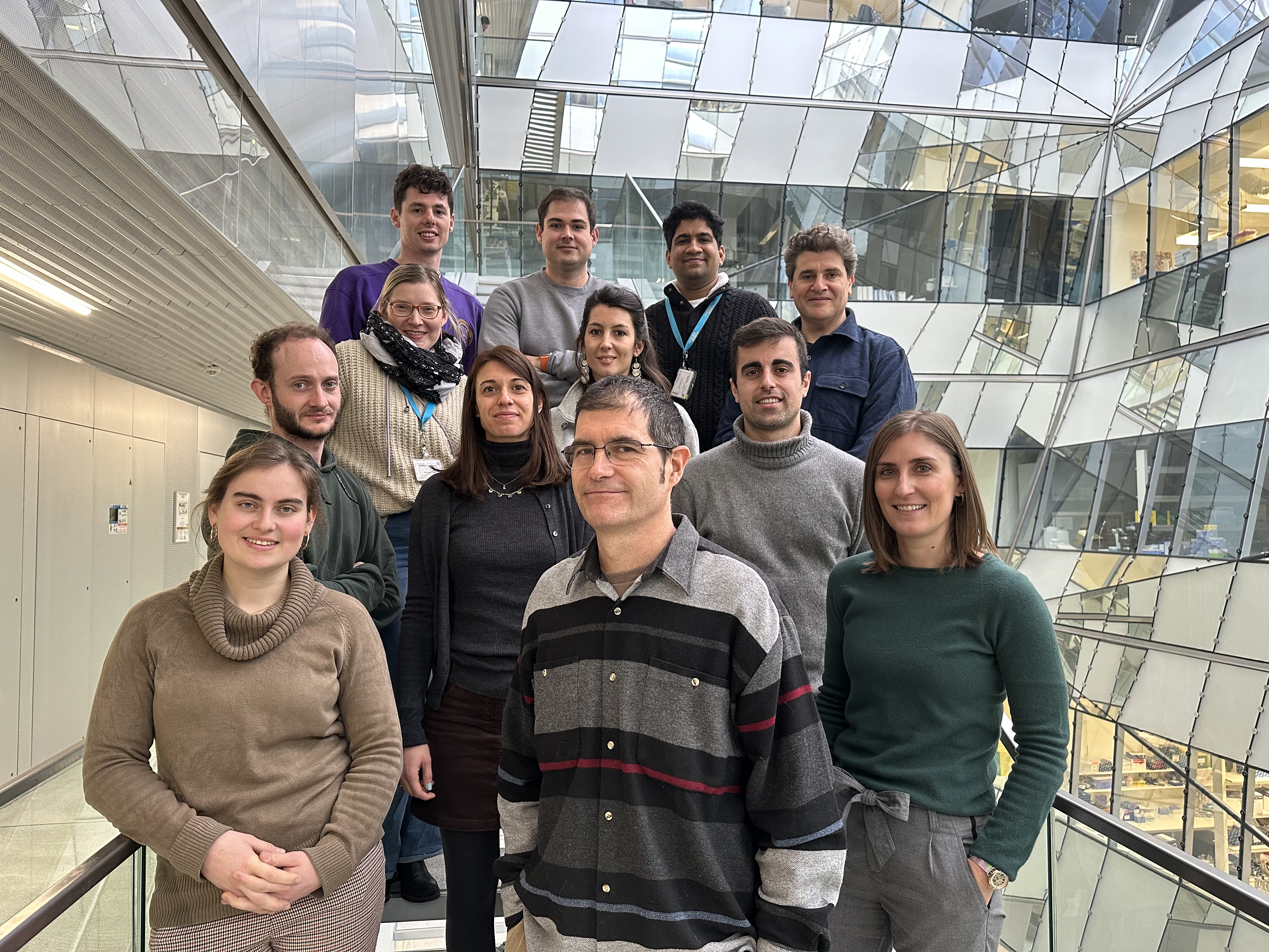
The team (January 2024)
Front row, from left to right: Domitille, David, Julie
Second row: Antoine, Sarka, Silvia, Lou, Unai
Third row: Paul, Vincent, Vivek, Etienne
General objective
Vesicular trafficking is one of the most salient features of synaptic physiology. In the tiny (less than 1 µm wide) chemical synapses, presynaptic vesicles concentrate and release neurotransmitter molecules which bind to post-synaptic receptors. The exocytosis and recycling of synaptic vesicles is a very prominent and essential feature of neuronal physiology that is highly controlled in time and space. Moreover, post-synaptic membrane trafficking, although not as prominent quantitatively, is pivotal for the maintenance of signal transduction complexes supporting synaptic transmission and plasticity. Most of our knowledge about synapse physiology comes from studying glutamatergic synapses which represent the majority of synapses in the brain. Nevertheless, other types of synapses, such as neuromodulatory dopaminergic synapses, could have a very different molecular composition and operate in a different way. However, because they represent a small minority of synapses formed from a very small number of neurons, their analysis has been difficult through classical cellular and molecular methods.
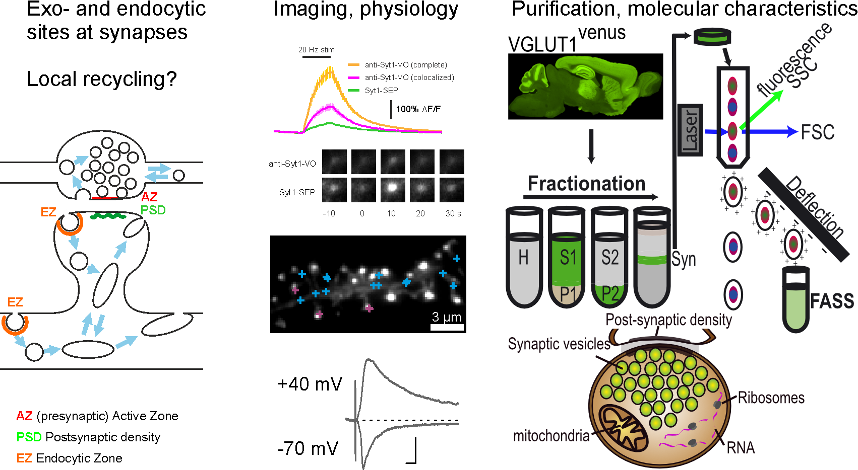
Our goal in the team is to use the most advanced fluorescence imaging techniques together with refined purification of synaptic elements (synaptosomes) to address the mechanisms regulating synapse function through membrane trafficking events in normal brain physiology or in the course of disease. To achieve this goal, we use, on top of the standard techniques of the modern neuroscience lab (molecular biology, biochemistry, imaging, electrophysiology), two unique expertise developed by the two PIs: first, with David Perrais, we develop methods to detect individual exocytosis and endocytosis events with pH sensitive fluorophores and perform quantitative imaging. Second, with Etienne Herzog, we purify synaptosomes from adult animals with fluorescence activated synaptosome sorting (FASS), which enables powerful proteomics, transcriptomics and functional approaches.
Altogether, we aim at identifying new pathways in specific synapses and test their relevance for synaptic nanostructure and function in the normal and diseased brain.
Expertise
News
Post-synaptic exocytosis and synaptic plasticity - Cell Reports Sept 2021
Synapses, the basic building blocks of neural networks, are both very stable and capable of rapid and long-lasting modifications, a phenomenon known as synaptic plasticity. The modification of a synapse often involves the addition of synaptic receptors (long-term potentiation or LTP) or the removal of part of the synaptic receptors (long-term depression or LTD). This rapid plasticity is possible because synaptic receptors are not immobile in the synapse but travel to intracellular compartments called recycling endosomes (RE). The regulation of RE trafficking has thus become an important topic of study for understanding the mechanisms of synaptic plasticity.
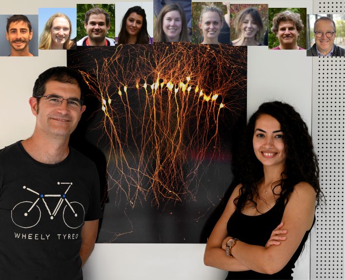
Picture of all contributors of this study. David Perrais and May Bakr stand in front of "The paint pipette", taken by May, which was awarded the best IINS picture prize in 2020.
In this study, we searched for the molecules involved in these phenomena, in particular the proteins responsible for exocytosis called SNAREs. The VAMP2 protein, target of the tetanus toxin (released by the bacterium responsible for tetanus and one of the most deadly in humans), was known to block LTP. However, to our surprise, it only marginally affected RE exocytosis. We therefore searched for other SNARE proteins and found VAMP4 to be responsible for the majority of RE exocytosis, whereas VAMP2 is involved in only a small fraction of exocytosis, but plays a major role in the exocytosis of REs containing AMPA-type postsynaptic receptors (see Figure). Furthermore, VAMP4 deletion also alters the trafficking of AMPA receptors that are in greater quantity at the surface of neurons, increasing synaptic transmission and limiting its plasticity by occlusion.
This work shows the great diversity of membrane trafficking mechanisms in the dendrites of neurons that allows receptors to be delivered when and where they are needed to regulate individual synapses. It was the result of a long-term work, over more than eight years, of many students, engineers and researchers of the IINS.
The team received an Equipe FRM award for their "SpaceNeuroModule" project
Spatio-temporal control of catecholamine neuromodulation of hippocampal pyramidal cells: from mechanisms of neuromodulator release to receptor signaling and trafficking (SpaceNeuroModule)
Scientific abstract
Chemical neurotransmission has been classified into fast neurotransmission and neuromodulation. Fast chemical neurotransmission, used by a majority of neurons in the central nervous system, is spatially restricted, thanks to a tight spatial control of neurotransmitter release at the active zone, the presence of cognate post-synaptic receptors and powerful recapture systems. In contrast, neuromodulation arises from a small number of neurons mediating slow communication to widespread neuronal networks. The sparsity of neuromodulatory synapses and the absence of direct ionotropic receptor signaling hampered our ability to resolve neuromodulation mechanisms at the micrometer and millisecond scale. In the project SpaceNeuroModule, we will address this question at noradrenaline projections in the hippocampus. We will use state-of-the art synaptosome sorting, proteomics, correlative cryo light and electron tomography (cryoCLEM), receptor interactome profiling, live cell fluorescence imaging and electrophysiology in vitro and ex vivo. We have defined 2 work packages to address the release of neuromodulators and the activation of their receptors at pyramidal dendrites, which regulates attention related learning and stress-related behaviors. First, we will determine the organization and dynamics of noradrenaline release sites. Second, we will assess the spatio-temporal extent of beta-adrenergic signaling in post-synaptic compartments. This research program will enable the formulation of design principles for neuromodulatory systems which will be tested further in disease models.
Lay abstract
The brain is composed of neurons which connect each other through synapses. A neuron sends neurotransmitter molecules to a connected neuron which captures them with specific receptors. Because a neuron receives up to thousands of synapses, some using the same neurotransmitter, each message must be sent precisely at the right place and travel minimal distance to the connected neuron and not spill over to neighboring synapses. Classical synapses are very specialized in ensuring this specific transmission with precise locations for release sites and receptors: decades of intense work from researchers around the world have discovered many of the building blocks of these synapses. A special category of neurons called neuromodulatory neurons, which comprise in particular dopaminergic neurons which degenerate in patients suffering from Parkinson’s disease, act in a different way. They do not mediate a fast signal but modulate large assemblies of neurons, even though they send molecules, in this case dopamine, to other neurons from specific sites. Because of technical difficulties such as the rarity of these neurons and their lack of fast signaling, we do not yet know how exactly is dopamine released and if specific sites are privileged, nor how receptors which are activated locally can convey the signal to the rest of the neuron. We will use state-of-the-art methods to purify neuromodulator synapses and identify new specific molecules (proteins). We will watch the structure of these sites with high resolution microscopes to understand what are the differences with classical synapses. Finally, we will image in brain tissue the release of neuromodulator and the activation and movement of their receptors to define how neuromodulation is organized and controlled in the brain.
Selected Publications
MORE
MORE
MORE
MORE
MORE
MORE
MORE
MORE
MORE
MORE
Members
« Researcher »
| HERZOG Etienne | Researcher | etienne.herzog@u-bordeaux.fr | +33533514861 |  |
| PERRAIS David | Researcher | david.perrais@u-bordeaux.fr | +33533514861 |  |
« Technical Staff »
| ANGIBAUD Julie | Technical staff | julie.angibaud@u-bordeaux.fr | +33533514748 |  |
| BOUIT Lou | Technical staff | lou.bouit.1@u-bordeaux.fr | +33533514861 |  |
| FIGUEIRA Michel | Technical staff | michel.figueira@u-bordeaux.fr | +33533514700 | |
| SCHWAB Lise | Technical staff | lise.schwab@u-bordeaux.fr | +33533514700 |  |
« Postdoc »
| JELINKOVA Sarka | Postdoc | sarka.jelinkova@u-bordeaux.fr | +33533514861 |  |
| LAPIOS Paul | Postdoc | paul.lapios@u-bordeaux.fr | +33533514779 |  |
| REYNAUD Antoine | Postdoc | antoine.reynaud@u-bordeaux.fr | +33533514700 | |
| SPOSINI Silvia | Postdoc | silvia.sposini@u-bordeaux.fr | +33533514861 |  |
« PhD student »
| DES ROBERT Domitille | PhD student | domitille.des-robert@u-bordeaux.fr | +33533514700 |  |
| DUFAU Audrey | PhD student | audrey.dufau@u-bordeaux.fr | +33533514700 |
« Student »
| CARLA LE BASTARD Anna | Student | anna.le-bastard@etu.u-bordeaux.fr | +33533514700 | |
| HOSPITAL Solène | Student | solene.hospital@etu.u-bordeaux.fr | +33533514700 | |
| MALI Aditi | Student | aditi.mali@u-bordeaux.fr | +33533514700 |
« Alumni & Guests »
May Bakr (PhD student 2017-2021 - Now Assistant Professor at American University of Cairo, Egypt)
Florencia Angelo (Lab Manager, 2015-2020 - Now Lab Manager at NutriNeuro Lab, Bordeaux, France)
Magalie Martineau (post-doc, 2015-2019 - Now Research and Development specialist at EIF Innovation, Paris, France)
Léa Claverie (PhD student, 2015-2019 - Now Project Manager at Euroquality, Bordeaux, France)
Domenico Azarnia Tehran (Guest, 2019)
Anne-Sophie Hafner (Guest, 2017-2018)
Thi Nhu Ngoc Van (post-doc, 2014-2016 - Now Project Manager at Sys2dia, Montpellier, France)
Julia Krapivkina (PhD student, 2012-2016 - Now Project Manager at Assystem, Paris, France)
Xiao Min Zhang (PhD Student, 2012-2016 - Now Assistant Professor at Sun Yat Sen University - China)
Morgane Rosendale (PhD student and post-doc, 2011-2016 - Now post-doctoral fellow at Institute for Molecular Sciences, Bordeaux)
Damien Jullié (PhD student, 2008-2012 - Now Research Specialist at UCSF Weill Institute for Neurosciences, San Francisco, USA)