Olivier Thoumine
Teams Cell biology Quantitative imaging of the cell
Researcher | PhD (DR) | CNRS
After completing an engineering degree at Ecole Centrale Paris, I carried out my Ph.D. at Georgia Tech (Atlanta), where I studied integrin-dependent mechanotransduction in the response of endothelial cells to hemodynamic forces. During my post-docs at Institut Curie (Paris) and Ecole Polytechnique Fédérale (Lausanne), I designed micromanipulation methods to precisely quantify the response of cells to mechanical deformations. After my recruitment by the CNRS in the team of Daniel Choquet (Bordeaux), I developed biomimetic systems coupled with high resolution imaging and predictive biophysical models, to probe the role of the cytoskeleton and adhesion proteins in growth cone motility and synaptogenesis. Then, I led an independent team at the Interdisciplinary Institute for Neuroscience from 2010 to 2023, focusing on the dynamics, localization, and function of synaptic adhesion molecules. In 2013, I spent a sabbatical year at UC Berkeley, learning optogenetics. In 2024, I joined the Team Quantitative Imaging of the Cell, where I develop projects around multimodal super-resolution imaging of synapses in 3D neuronal samples.
To probe the ability of adhesion molecules to promote neuronal development, we developed a biomimetic system consisting of culturing primary dissociated neurons on glass substrates micropatterned with purified Fc-tagged adhesion molecules (N-cadherin, Neurexin-1β, SynCAM1). Using this high-content device, we showed that N-cadherin elicits axonal growth through a clutch mechanism (Garcia et al. PNAS 2015), while SynCAM1 and Neurexin-1β induce pre- and post-synaptic differentiation, respectively (Czöndör et al. Nat Commun 2013; Czöndör et al. Meth Cell Biol 2025) (Fig. 1).
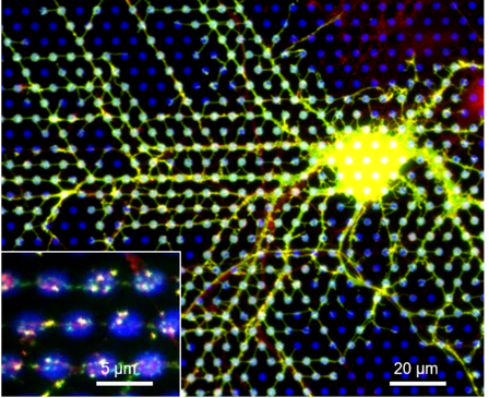
Figure 1. Hippocampal neuron expressing neuroligin-1 and PSD-95-GFP (green) cultured on micropatterned substrates coated with neurexin1β-Fc (blue dots). The inset shows the formation of PSD-95-GFP and AMPA receptor clusters at neurexin1β-Fc coated dots.
To accompany the progress in fluorescence live-cell and super-resolution microscopy, we developed a real-time simulator of single molecule dynamics called FluoSim (Lagardère et al., Sci Rep 2020), which can reproduce a variety of imaging experiments performed on membrane proteins (Fig. 2). FluoSim was validated against SPT, PAF, FRAP, FCS, and STORM data obtained on the canonical neurexin-neuroligin complex, using a small number of kinetic and photophysical parameters. This software is an interesting tool to improve experimental design and analysis, as well as to train users on the most challenging imaging methods.
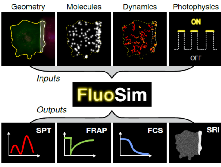
Figure 2. General principle of FluoSim
A decade ago, we provided evidence that neuroligin-1 was a ligand-activated adhesion molecule, i.e. binding to pre-synaptic neurexin-1β was able to switch the affinity of neuroligin-1 towards post-synaptic scaffolding proteins (PSD-95 versus gephyrin), this process being regulated by the phosphorylation of a unique intracellular tyrosine (Y782) (Giannone et al., Cell Rep 2013). Using two tyrosine point mutants (Y782A/F), we further showed that neuroligin-1 phosphorylation was implicated in AMPA receptor synaptic recruitment and long term potentiation (LTP), and identified the implicated tyrosine kinase receptors (i.e. Trks) (Letellier et al., Nat Commun 2018; Sziber et al., Frontiers Mol Neurosci. 2024). Finally, we optogenetically triggered the phosphorylation of native neuroligin-1 using a light-gated tyrosine kinase (FGF receptor 1), allowing a control of post-synaptic differentiation without affecting neuroligin-1 expression level (Letellier et al., eLife 2020).
By developing specific labeling strategies relying on small monomeric probes combined with super-resolution fluorescence imaging, we tracked individual β-neurexin-1, neuroligin-1, and MAM-domain GPI-Anchored (MDGAs) molecules. We showed that both β-neurexin-1 and neuroligin-1 reach synapses through a diffusion-trap mechanism and form synaptic nanodomains (Chamma et al. Nat Commun 2016; Lagardère et al. Front Synaptic Neurosci 2021) (Fig. 3), while MDGAs that bind neuroligins in cis and prevent neurexin binding, display fast membrane diffusion and do not accumulate at synapses (Toledo et al. eLife 2022).
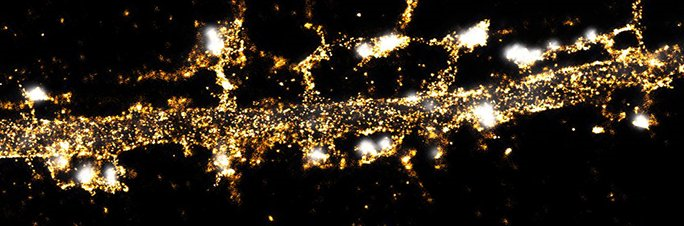
Figure 3. Single molecule localization map of biotinylated recombinant bAP-neuroligin-1 labeled with Alexa647-conjugated monomeric streptavidin, showing a homogeneous distribution in the dendritic shaft and accumulation at post-synaptic densities (labeled with the Homer1c-GFP reporter in white).
To study the localization of endogenous neuroligin-1, we generated a transgenic mouse strain in which neuroligin-1 carries a small N-terminal biotin acceptor peptide (bAP) tag that can be enzymatically biotinylated, followed by selective isolation or visualization using high-affinity streptavidin conjugates (Ducrot et al. PNAS 2025). We show that bAP-neuroligin-1 binds PSD-95 and gephyrin and populates both excitatory and inhibitory synapses, challenging the historical view that neuroligin-1 is exclusively localized at excitatory synapses. Using super-resolution optical and electron microscopy, we further highlight that synaptic bAP-neuroligin-1 forms a subset of nanodomains, which contain each a few neuroligin-1 dimers and whose number positively scales with the post-synapse size.
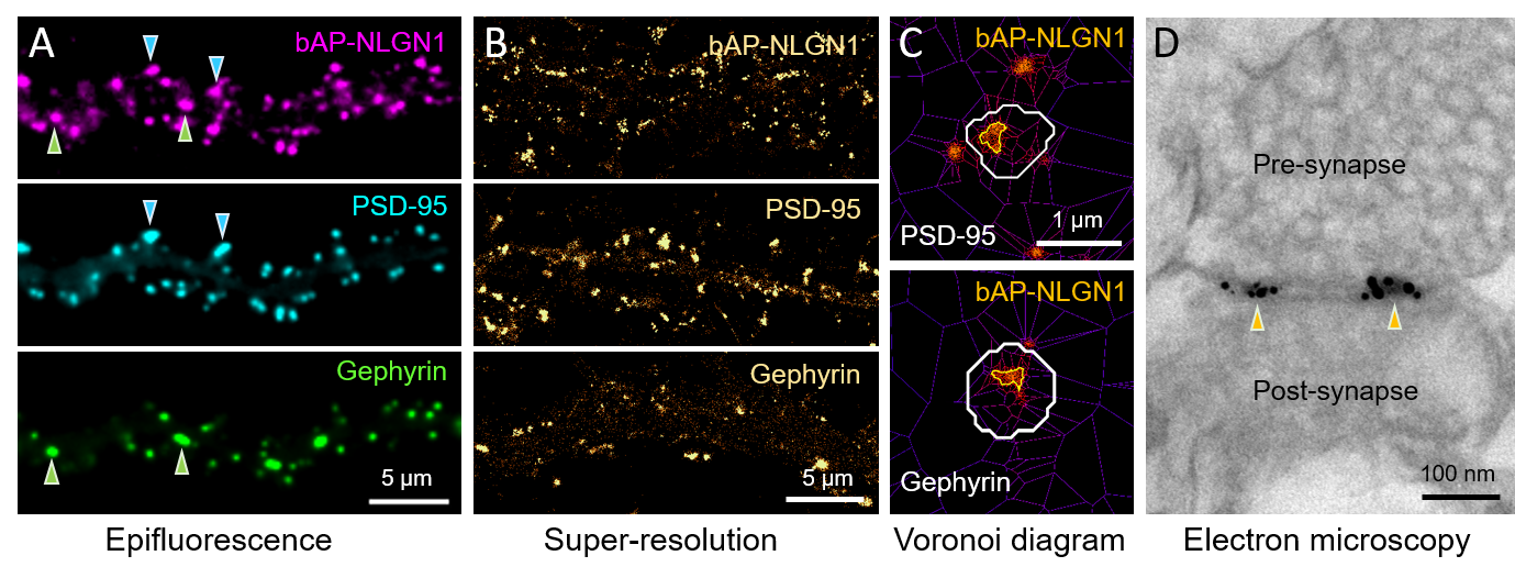
Figure 4. Endogenous neuroligin1 at excitatory & inhibitory synapses. (A) Epifluorescence images of biotinylated bAP-NLGN1 stained with streptavidin-AF647, PSD-95 and gephyrin being labeled with intrabodies fused to mRuby and GFP. (B) dSTORM on bAP-NLGN1 and PALM on PSD-95 and gephyrin. (C) NLGN1 forms a subset of nanodomains within excitatory and inhibitory synapses. (D) Electron micrograph of the synaptic cleft showing two bAP-NLGN1 nanodomains labeled with streptavidin-Nanogold and enhanced with silver.
Achievements
Micropatterning assay to screen for synapse differentiation
To probe the ability of adhesion molecules to promote neuronal development, we developed a biomimetic system consisting of culturing primary dissociated neurons on glass substrates micropatterned with purified Fc-tagged adhesion molecules (N-cadherin, Neurexin-1β, SynCAM1). Using this high-content device, we showed that N-cadherin elicits axonal growth through a clutch mechanism (Garcia et al. PNAS 2015), while SynCAM1 and Neurexin-1β induce pre- and post-synaptic differentiation, respectively (Czöndör et al. Nat Commun 2013; Czöndör et al. Meth Cell Biol 2025) (Fig. 1).
Figure 1. Hippocampal neuron expressing neuroligin-1 and PSD-95-GFP (green) cultured on micropatterned substrates coated with neurexin1β-Fc (blue dots). The inset shows the formation of PSD-95-GFP and AMPA receptor clusters at neurexin1β-Fc coated dots.
Computer simulations based on single molecule dynamics to quantitatively interpret fluorescence microscopy experiments
To accompany the progress in fluorescence live-cell and super-resolution microscopy, we developed a real-time simulator of single molecule dynamics called FluoSim (Lagardère et al., Sci Rep 2020), which can reproduce a variety of imaging experiments performed on membrane proteins (Fig. 2). FluoSim was validated against SPT, PAF, FRAP, FCS, and STORM data obtained on the canonical neurexin-neuroligin complex, using a small number of kinetic and photophysical parameters. This software is an interesting tool to improve experimental design and analysis, as well as to train users on the most challenging imaging methods.
Figure 2. General principle of FluoSim
Role of neuroligin-1 in post-synaptic differentition
A decade ago, we provided evidence that neuroligin-1 was a ligand-activated adhesion molecule, i.e. binding to pre-synaptic neurexin-1β was able to switch the affinity of neuroligin-1 towards post-synaptic scaffolding proteins (PSD-95 versus gephyrin), this process being regulated by the phosphorylation of a unique intracellular tyrosine (Y782) (Giannone et al., Cell Rep 2013). Using two tyrosine point mutants (Y782A/F), we further showed that neuroligin-1 phosphorylation was implicated in AMPA receptor synaptic recruitment and long term potentiation (LTP), and identified the implicated tyrosine kinase receptors (i.e. Trks) (Letellier et al., Nat Commun 2018; Sziber et al., Frontiers Mol Neurosci. 2024). Finally, we optogenetically triggered the phosphorylation of native neuroligin-1 using a light-gated tyrosine kinase (FGF receptor 1), allowing a control of post-synaptic differentiation without affecting neuroligin-1 expression level (Letellier et al., eLife 2020).
Dynamics and localization of recombinant neuroligin-1 at synapses
By developing specific labeling strategies relying on small monomeric probes combined with super-resolution fluorescence imaging, we tracked individual β-neurexin-1, neuroligin-1, and MAM-domain GPI-Anchored (MDGAs) molecules. We showed that both β-neurexin-1 and neuroligin-1 reach synapses through a diffusion-trap mechanism and form synaptic nanodomains (Chamma et al. Nat Commun 2016; Lagardère et al. Front Synaptic Neurosci 2021) (Fig. 3), while MDGAs that bind neuroligins in cis and prevent neurexin binding, display fast membrane diffusion and do not accumulate at synapses (Toledo et al. eLife 2022).
Figure 3. Single molecule localization map of biotinylated recombinant bAP-neuroligin-1 labeled with Alexa647-conjugated monomeric streptavidin, showing a homogeneous distribution in the dendritic shaft and accumulation at post-synaptic densities (labeled with the Homer1c-GFP reporter in white).
Localization of endogenous neuroligin-1 at excitatory and inhibitory synapses
To study the localization of endogenous neuroligin-1, we generated a transgenic mouse strain in which neuroligin-1 carries a small N-terminal biotin acceptor peptide (bAP) tag that can be enzymatically biotinylated, followed by selective isolation or visualization using high-affinity streptavidin conjugates (Ducrot et al. PNAS 2025). We show that bAP-neuroligin-1 binds PSD-95 and gephyrin and populates both excitatory and inhibitory synapses, challenging the historical view that neuroligin-1 is exclusively localized at excitatory synapses. Using super-resolution optical and electron microscopy, we further highlight that synaptic bAP-neuroligin-1 forms a subset of nanodomains, which contain each a few neuroligin-1 dimers and whose number positively scales with the post-synapse size.
Figure 4. Endogenous neuroligin1 at excitatory & inhibitory synapses. (A) Epifluorescence images of biotinylated bAP-NLGN1 stained with streptavidin-AF647, PSD-95 and gephyrin being labeled with intrabodies fused to mRuby and GFP. (B) dSTORM on bAP-NLGN1 and PALM on PSD-95 and gephyrin. (C) NLGN1 forms a subset of nanodomains within excitatory and inhibitory synapses. (D) Electron micrograph of the synaptic cleft showing two bAP-NLGN1 nanodomains labeled with streptavidin-Nanogold and enhanced with silver.